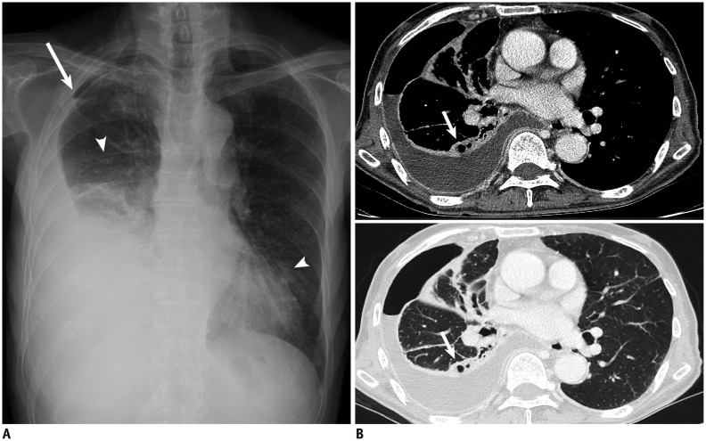
Fig. 1. Chest radiograph and CT images obtained from 86-year-old man with pulmonary disease and hydropneumothorax caused by Mycobacterium avium (Case 4).
A. Chest radiography shows hydropneumothorax (arrow) in right hemithorax. Small cavitary or non-cavitary nodules and branching nodular structures (tree-in-bud pattern) (arrowheads) are seen in both lungs. B. CT images with lung and mediastinal window setting demonstrate hydropneumothorax and enhancing pleural thickening suggesting pleural empyema. Note broncho-pleural fistulas (arrow) that developed from prior cavitary lesion (not shown) in same area.

