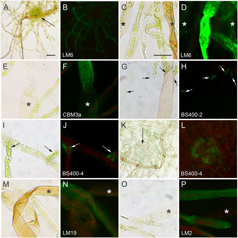
FIGURE 4.
Physcomitrella patens rhizoids cultured on medium containing auxin (1 μM naphthalene acetic acid) for 2 weeks, fixed and labeled with antibodies or CBMs. Rhizoids were imaged with bright field (A,C,E,G,I,K,M,O) and fluorescence optics (B,D,F,H,J,L,N,P) after labeling with (A–D) anti-1,5-α-L-arabinan (LM6), (E,F) CBM3a for crystalline cellulose, (G,H) anti-callose (BS-400-2), which labels cross walls indicated by arrows, (I–L) anti-mannan (BS-400-4), which labels cell junctions indicated by arrows, (M,N) anti-HG (LM19), and (O,P) anti-arabinogalactan protein (LM2). Mature pigmented rhizoids are indicated with asterisk. Bar = 200 μm in (A) for (A,B). Bar = 100 μm in (C) for (C–P).

