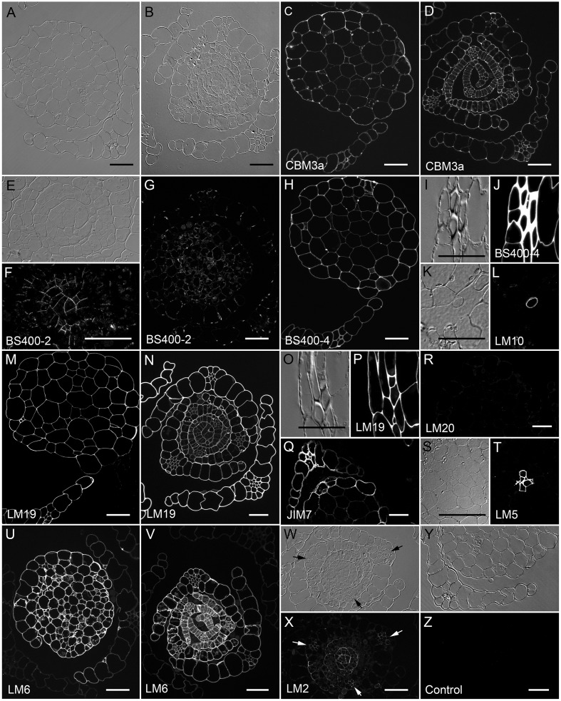
FIGURE 5.
Physcomitrella patens gametophores labeled with antibodies or CBMs. Transverse sections through mature stems and leaves (A,C,G,H,M,Q,R,Y,Z) and meristems and young leaves (B,D,E,F,N,V,W,X) imaged with DIC optics (A,B,E,I,K,O,S,W,Y) and with fluorescence optics after labeling for (C,D) crystalline cellulose with CBM3a, (F,G) callose with BS400-2, (H,J) mannan with BS400-4, (L) xylan with LM10, homogalacturonon with (M,N,P) LM19, (Q) JIM7 or (R) LM20, (T) 1,4-β-galactan with LM5, (U,V) 1,5-α-L-arabinan with LM6, (X) arabinogalactan proteins with LM2, or (Z) no primary antibody control. Higher magnification views show labeling of (E,F) cell plates with anti-callose, (I,J) the thick walls of stereid cells with anti-HG, (K,L) axillary hairs with anti-xylan, (O,P) the middle lamella of stereid cells with anti-homogalaturonon and (S,T) hydroids. All fluorescence images were captured using identical exposure conditions with the exceptions of (L,Q,R), which were captured using a different microscope. Bars = 50 μm.

