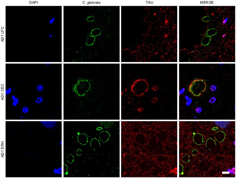
Figure 1.
Corpora amylacea are detected in different regions of the CNS. Tissue sections (5 μm) from different CNS regions of patient AD1 were tested as follows: lateral frontal cortex (LFC), cerebellar cortex (CEC) and entorhinal cortex/hippocampus (ERH). Immunohistochemistry analysis was carried out by double immunofluorescence staining employing a rabbit polyclonal anti-C. glabrata antibody (green) and a mouse monoclonal anti-tau antibody (red). Sections were mounted and observed by confocal microscopy after incubation with the corresponding secondary antibodies, as described in Materials and Methods. Overlapping red and green pixels appear as orange/yellow. DAPI appears in blue. Scale bar: 10 μm.

