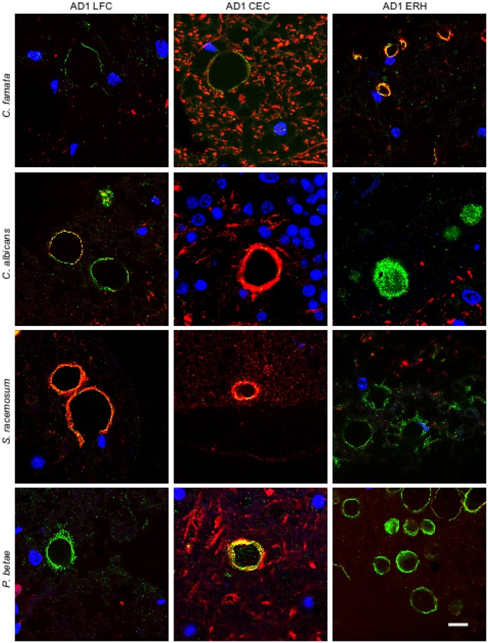
Figure 2.
Immunoreactivity of corpora amylacea against different antifungal antibodies. Tissue sections analyzed from patient AD1 are indicated in the figure. Immunohistochemistry analysis was carried out using a monoclonal antibody to human neurofilaments (red) and rabbit polyclonal antibodies raised against the following fungi: C. famata, C. albicans. S. racemosum, and P. betae (green). Sections were mounted and examined by confocal microscopy after incubation with the corresponding secondary antibodies. Orange/yellow corresponds to red and green pixels. DAPI (blue) and scale bar: 10 μm.

