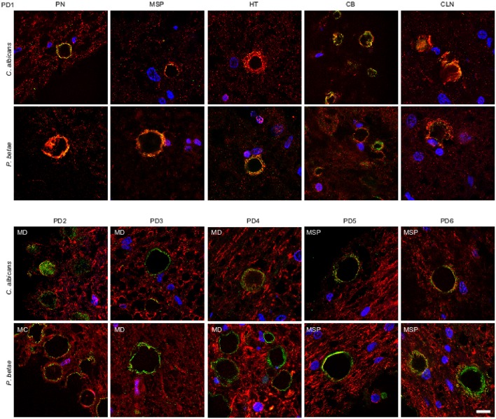
Figure 5.
Detection of fungal proteins in corpora amylacea from PD patients. Tissue sections of patient PD1 were obtained from the following regions: pons (PN), mesencephalon (MSP), hypothalamus (HT), callosal body (CB), and caudate and lenticular nuclei (CLN) (upper panels). Different regions of the CNS from five additional PD patients (PD2-PD6) were also analyzed (lower panels). Sections were incubated with rabbit polyclonal antibodies against C. albicans and P. betae (green) and a monoclonal antibody against α-tubulin (red). Sections were mounted and examined by confocal microscopy after incubation with the corresponding secondary antibodies. Overlapping red and green pixels appear as orange/yellow. DAPI and scale bar as in Figure 1.

