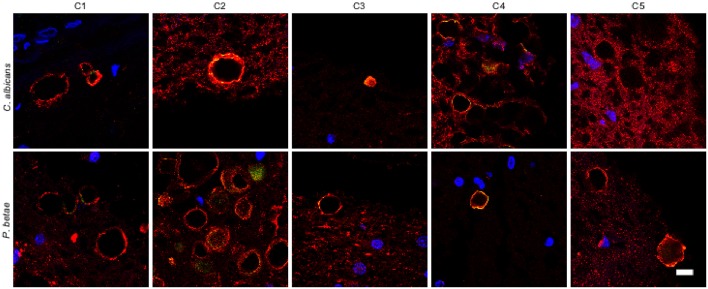
Figure 6.
Analysis of corpora amylacea present in the CNS of control individuals. Tissue sections from the ERH of five control individuals (C1–C5) were analyzed by immunohistochemistry. Sections were incubated with rabbit polyclonal antibodies against C. albicans and P. betae (green) and a monoclonal antibody against α-tubulin (red). Sections were mounted and examined by confocal microscopy after incubation with the corresponding secondary antibodies. Overlapping red and green pixels appear as orange/yellow. Note the near absence of activity against the fungal antibodies. DAPI and scale bar as in Figure 1.

