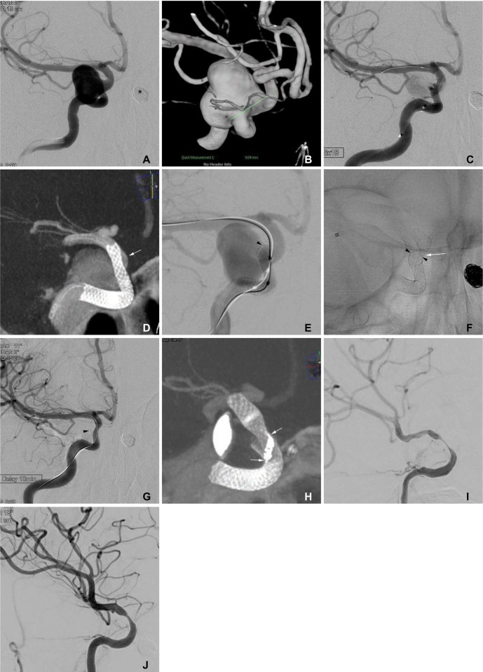
Fig. 1. A 51-year-old woman with bilateral distal internal carotid artery aneurysms.
A, B. Frontal projection and 3-D reconstruction images show a large aneurysm at the intradural para-ophthalmic segment of the right internal carotid artery. The asterisk indicates a coil-embolized aneurysm at the left distal internal carotid artery. The asterisk indicates the coil embolized aneurysm of left internal carotid artery. C. Angiogram after Pipeline implantation shows markedly decreased flow into the aneurysm sac. D. A flat-panel CT image shows incomplete expansion of the distal portion of the Pipeline resulting in poor wall apposition (arrow). E. After ballooning for wall apposition of the Pipeline, the distal end (arrowhead) of the pipeline was partially herniated into the aneurysm sac, resulting in alleviation of the flow diversion effect. F. A spot image after the second Pipeline implantation in a telescopic manner. Note the waist of the second Pipeline at the distal end (arrowhead) of the first Pipeline, which partially herniated into the sac. G. After using a balloon for the apposition of the second Pipeline, the aneurysm sac was no longer visualized. The arrowhead indicates the distal end partially herniated into the sac. H. A flat-panel CT image showing that a mild degree of the waist of the second pipeline still remained at the end of the first pipeline. Note the twisted struts (arrows) at the waist point. I. The 6-month follow-up angiogram shows a severe degree of in-stent stenosis, even though the patient remained asymptomatic. The left anterior cerebral artery was supplied via the anterior communicating artery from the left internal carotid artery (not shown). J. The 12-month follow-up angiogram shows improvement of the in-stent stenosis.

