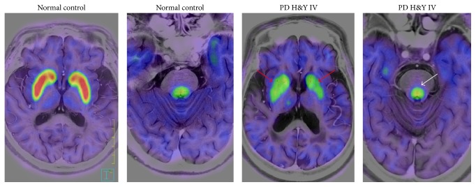
Figure 1.
Representative FMT-positron emission tomography images of a healthy individual and a patient with advanced stage PD. Decrease of FMT uptake is obvious in the putamen and caudate (red arrow) in the PD patient. FMT uptake in the locus coeruleus (white arrow) in this patient is relatively maintained. H&Y: Hoehn and Yahr stage.

