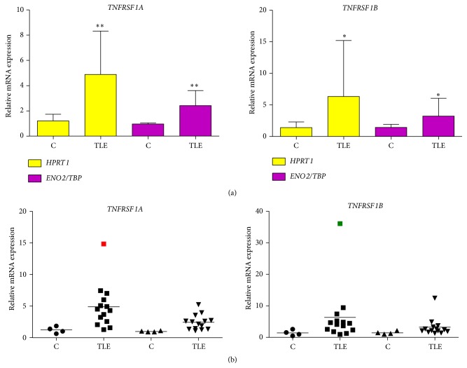
Figure 2.
Hippocampal gene expression of TNFRSF1A and TNFRSF1B in TLE(HS) patients versus post mortem controls. HPRT1 and ENO2/TBP were used as reference genes. One of the post mortem control samples was randomly chosen as the reference and its relative quantification was always 1.0. All quantification data for the remaining samples, including controls and patients, was benchmarked to the reference sample. Samples were separated in two groups: TLE(HS) patients (n = 14) and post mortem controls (n = 4). (a) Columns are means with SD. Mann-Whitney U tests were used for the comparison between groups. ∗ P < 0.05; ∗∗ P < 0.01. (b) Marks are different geometric figures, which represent the gene expression mean of the samples. The horizontal line is the mean of the group analyzed. Circles and squares correspond to gene expression with HPRT1 as the reference. The two kinds of triangles correspond to gene expression with ENO2/TBP as the reference. The red and green squares (outliers) correspond to TLE 09 and TLE 03 patients, respectively (see Table 1 in Teocchi et al., 2013 [8]).

