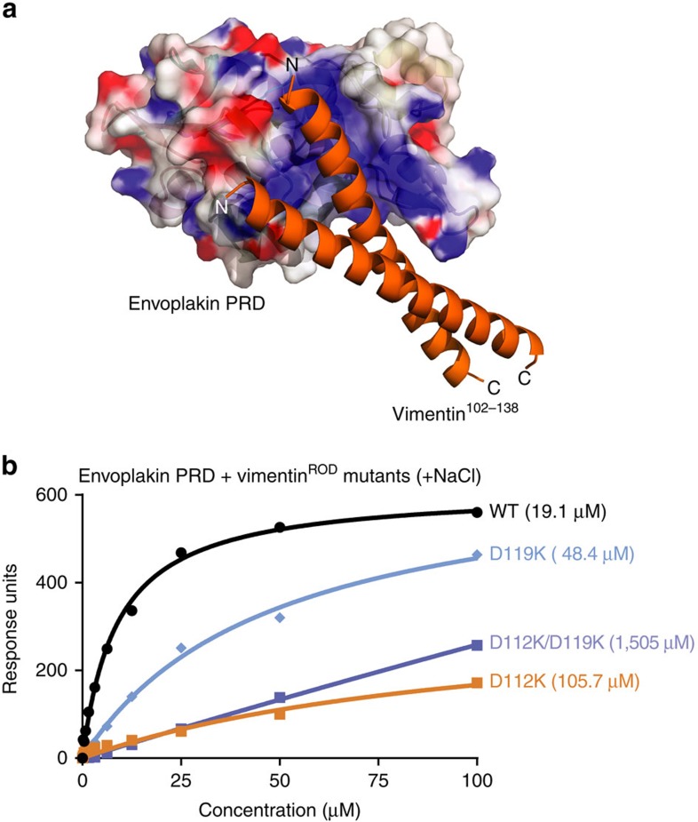
Figure 6. Modelling and binding studies of the envoplakin PRD–vimentin complex.
(a) The envoplakin PRD structure (coloured as in Fig. 3) was docked with vimentin Asn102-Leu138 (orange) (PDB 3G1E) using HADDOCK. (b) SPR analysis of wild-type envoplakin PRD binding to wild-type and mutant vimentinROD in the presence of 150 mM NaCl. The figures in parentheses are KD values in μM. The data shown is representative of a number of experiments. KD values were estimated to be: WT=19.1±1.3 μM, D112K=105.7±45.6 μM, D119K=48.4±5.5 μM and D112K/D119K=1505±139 μM.

