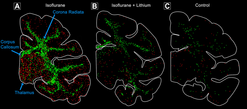
Figure 2. Pattern of neuronal and glial apoptosis following isoflurane anaesthesia in infant monkey at the level of the thalamus.
Computer generated duel plots show apoptotic (AC3-positive) neurons (red) and glia (green) in animals after a 5-hour isoflurane anaesthesia (A) 5 hours of isoflurane plus lithium co-treatment (B) or no anaesthesia (control; (C)). Isoflurane produces high levels of neuronal apoptosis in the thalamus and cortex whereas oligo apoptosis is concentrated in the white matter including the corpus callosum and corona radiata. Lithium co-treatment during isoflurane anaesthesia dramatically reduces apoptosis in both cell types similar to that seen in controls (no anaesthesia).

