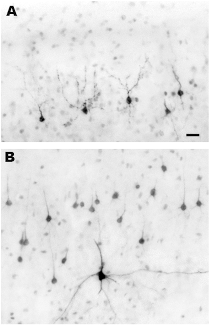
Figure 4. Histological sections from the primary visual cortex of an infant monkey exposed to ISO, illustrating the appearance of neuronal profiles that are stained by activated caspase 3 (AC3), a selective marker for apoptosis.

As shown in Fig. 2, the neurotoxic action of ISO selectively impinges on neurons in layers II and V of the primary visual cortex. The dark profiles in panel (A) are AC3-positive neurons undergoing apoptotic cell death in layer II, and those in panel (B) are AC3-positive neurons undergoing apoptotic cell death in layer V. The dying neurons in layer II panel (A) are GABAergic inhibitory neurons. Most of those in layer V panel (B) are small pyramidal neurons that are thought to be glutamatergic excitatory neurons. The single larger multipolar profile in panel (B) has the morphological characteristics of a Martinotti neuron35, which is believed to exert an inhibitory action. Bar in A = 20μm for both panels (A,B).
