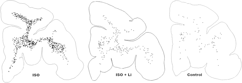
Figure 6. Pattern of oligo-apoptosis induced by isoflurane anaesthesia in the infant monkey brain.
Here we show computer plots at the level of the prefrontal cortex depicting the density and location of apoptotic oligos (black dots) under each treatment condition. The apoptotic oligo profiles are confined to a white matter distribution under each treatment condition, but are present in high density in the ISO-exposed brain, compared to lower and lowest density in the ISO + Li and control brains, respectively. The distribution of degenerating oligos shown here is representative of the distribution throughout the brain; they are diffusely and relatively evenly distributed throughout all white matter pathways.

