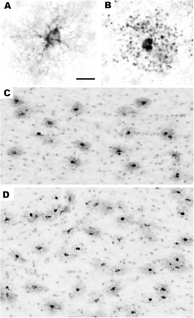
Figure 7. Histological sections illustrating the appearance of degenerating oligos stained by AC3 or myelin basic protein (MBP).

We have shown9,11 that the white matter cells that undergo apoptosis when exposed to isoflurane co-label for AC3 (apoptosis marker) and MBP. This identifies these cells as oligodendrocytes (oligos) because oligos are the only cell type in the brain that contains MBP. Panels (A,B) are both stained with antibodies to MBP. A illustrates the appearance of a normal healthy oligo for comparison with the cell in B that is in an advanced stage of apoptosis following exposure to isoflurane. As the oligo degenerates, its processes become fragmented into many small dark balls that distribute around the cell body in a star burst configuration (B). When oligos undergo apoptosis they stain selectively and intensely with antibodies to AC3. Therefore, this is the method we employ for mapping and quantifying apoptotic oligos under various treatment conditions. Panels (C,D) illustrate the appearance of apoptotic oligos following isoflurane exposure in two major white matter tracts, the corpus callosum (C) and internal capsule (D). In each of these white matter pathways apoptotic oligos stand out as dark profiles which are typically surrounded by a smudgy circle of tissue discoloration which reflects leakage of AC3 into the neuropil from their disintegrating processes. Bar in A = 15 μm in A and B, and = 75 μm in C and 100 μm in (D).
