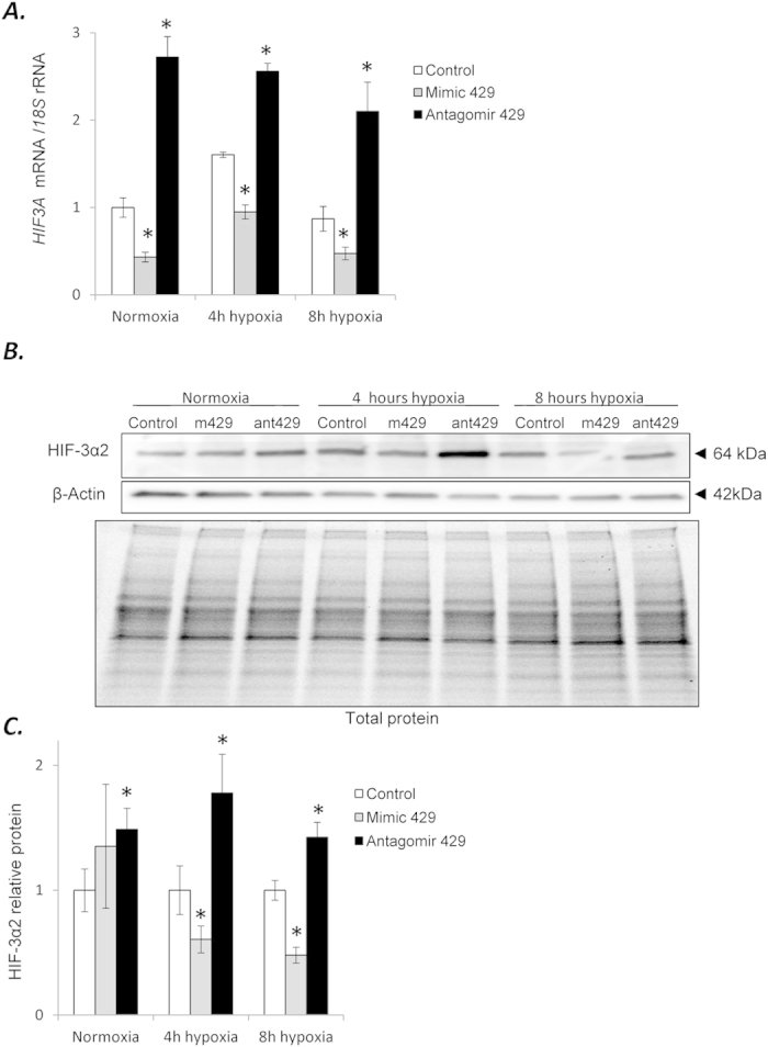
Figure 3.

(A) miR-429 alters the expression of HIF3A. HUVECs were transfected with miR-429 mimic or antagomir, and the mRNA levels were monitored in normoxic conditions and after 4 h and 8 h of exposure to hypoxia. HIF3A mRNA levels from 3 independent experiments (n = 12) are plotted normalized to 18S rRNA levels and expressed as a fold change over the transfection control. Significant changes (p < 0.05) are marked with an “*”. (B) The corresponding changes of HIF-3α2 protein levels of were detected with SDS-PAGE and Western Blot and normalized to the β-actin and total protein levels. (C) The bar graphs show the relative protein amounts at each condition normalized to the transfection control. (D) 2 individual samples (4 μg of total protein per lane) were tested for each treatment and the experiments were repeated twice. Mimic 429 (m429) and antagomir 429 (ant429) are shown for each condition.
