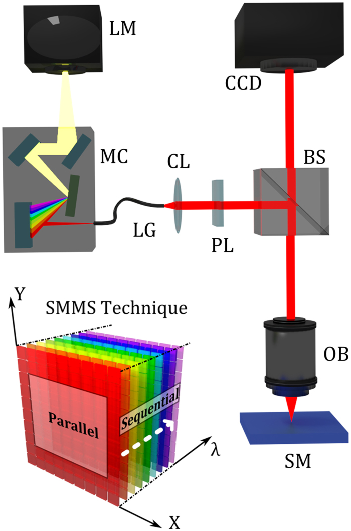
Figure 1. Working principle and practical realization of the SMMS technique in reflection mode configuration.

The white light from a lamp (LM) is directed to a motorized monochromator (MC) that disperses the light into its constituent wavelengths. A narrow band of the dispersed spectrum passing from the exit slit of the monochromator is directed to a collimator (CL) and a linear polarizer (PL) through a light guide (LG). Thus, monochromatic light is directed and focused to the sample (SM) by a beam splitter (BS) and a dark-field objective (OB). A high resolution CCD camera placed at the image plane of the experimental setup collects, for each illumination wavelength, λ, the scattered light coming from the full sample surface area. The SMMS technique can be employed also as a standard optical microscope by making use of the zero-order diffraction of the monochromator grating. The left bottom inset is a schematic drawing of the SMMS working principle; the measurements are acquired in parallel (multiplexed) for the spatial coordinates X and Y, and sequentially along the spectral coordinate λ.
