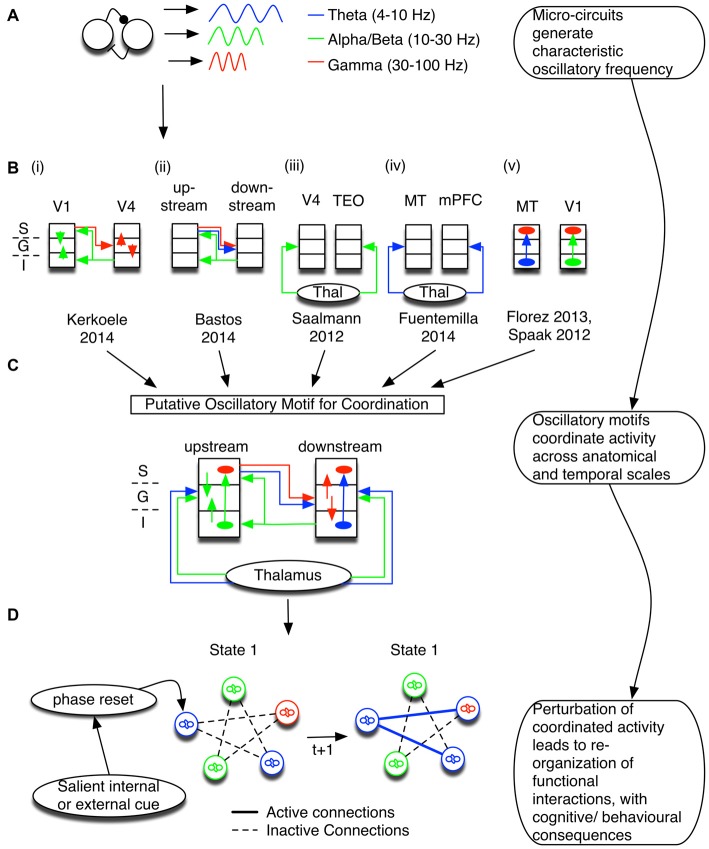
Figure 1.
Coordination of information flow is maintained by oscillatory dynamic circuit motifs. (A) Canonical circuit motifs give rise to frequency-specific oscillations, such as theta (blue), alpha and/or beta (green), and gamma (red). (B) These motifs are embedded within cortical micro-columns and support information processing between layers and cortical areas. Summarized here are the results of six studies that suggest specific relationships of oscillatory activation signature and anatomical circuit structure. The left most inset depicts the cortical layers S, G, and I, corresponding to the supra-granular, granular, and infragranular layers, respectively. (i,ii) In visual cortex, gamma activity follows feedforward connections (red; van Kerkoerle et al., 2014; Bastos et al., 2015) and co-occurs with low frequency theta (Bastos et al., 2015). On the other hand, alpha/low beta activity correlates with the feedback direction (green; Bastos et al., 2015). (iii,iv) The entrainment of cortical oscillations may depend on thalamic input at theta, alpha and/or beta band frequencies (Saalmann et al., 2012; Fuentemilla et al., 2014). (v) Long-distance oscillatory coordination can then affect processing within a micro-column; for example, theta activity and alpha generated in deep layers modulated gamma activity in superficial layers (Spaak et al., 2012; Florez et al., 2013). (C) The six studies outlined in (B) have been combined to highlight a putative oscillatory motif coordinating distant sites via interactions between different frequencies. The putative combined motif suggests that a mixture of oscillatory dynamic circuit motifs coordinates information processing between cortical laminae and across brain areas. Different circuit motifs are responsible for generating/propagating specific oscillations, and are recruited to fulfill specific functions. Note that this conceptual model is dominated by studies of visual processing. We stress that the purpose of combining these studies is to set out a framework to understand how local and distant circuits may functionally interact. Thus, it is likely that brain areas with different laminar structures have a different coordination profile, and future studies across multiple brain areas are necessary to test the predictions of such a framework (see Box 1). (D) Not only are oscillations constrained by anatomy, but oscillatory networks also emerge in relation to specific task contexts. Tracking changes in the phase of interacting oscillatory sources can thus be used to make predictions about anatomical and neurophysiological mechanisms underlying functional changes. Abbrevations: TEO, temporo-occipital; MT, medial temporal; mPFC, medial prefrontal cortex.

