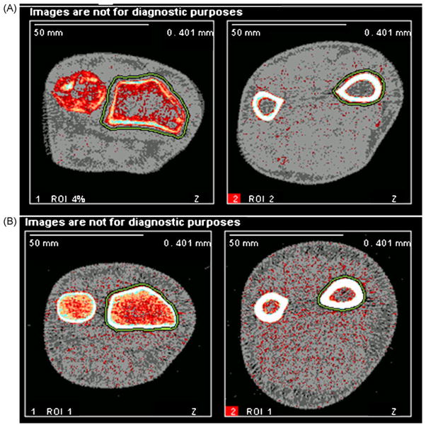
Fig. 1.
pQCT images of hERKO subject and normal man. This figure depicts pQCT images at the 4% (left) and 20% (right) distal radius in the hERKO subject (top row, A) and a man with similar arm length (bottom row, B). Decreased cortical thickness is apparent at both sites, as well as increased periosteal circumference and decreased trabecular vBMD, at the 4% distal radius.

