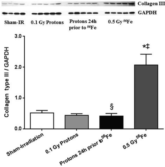
Figure 2.
Increased cardiac levels of collagen type III (quantification of the 75 kDa fragment as indicated in Supplementary Fig. 1) was observed at 7 days after exposure to 56Fe. A priming dose of protons prevented the increase in collagen type III in response to 56Fe exposure. Western-blot image indicates 4-5 biological repeats in each radiation group. Graphs indicate average ± SEM, n=4-5. Quantification of collagen type III divided by GAPDH for sham-irradiation: 0.51 ± 0.08; for 0.1 Gy protons: 0.43 ± 0.06; for 0.5 Gy 56Fe: 2.07 ± 0.34; for the consecutive exposure group: 0.41 ± 0.09. *p<0.05 compared to sham-irradiated control, ‡p<0.05 compared to proton exposure, §p<0.05 compared to 56Fe exposure.

