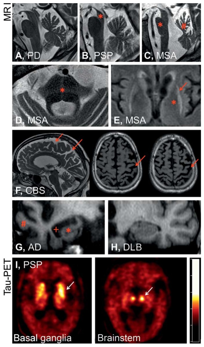
eFigure.
Magnetic resonance imaging shows in the midsagittal plane no specific atrophy pattern for Parkinson’s disease (PD) (A), whereas in progressive supranuclear palsy (PSP) (B), midbrain atrophy (*) is visible, and for multiple system atrophy (MSA) (C) the image shows pontine atrophy (*) and atrophy in the upper portion of the cerebellar vermis (#). Furthermore, signal anomalies in T2 weighted MRI with 1.5 Tesla are suggestive of, but not highly sensitive for, MSA—especially a cross-shaped hypointensity in the pons (hot cross bun sign, *, D) and a hypointense putamen (*) with hyperintense putaminal rim (putaminal rim sign, arrow, E). In corticobasal syndrome (CBS) (F), what is typically seen is asymmetric parietal atrophy with atrophy of the gyri and accentuation of the sulci (arrows). In the coronal plane, atrophy of the hippocampus (*) and the temporal lobe (#) with dilatation of the temporal horns of the lateral ventricles (+) are typically seen in Alzheimer’s dementia (AD). However, findings are normal in dementia with Lewy bodies (DLB) (H). Molecular diagnosis of these disorders is the objective of current research, such as has been shown in the example of a patient with PSP by confirming tau deposition in the basal ganglia and the brainstem on positron emission tomography (PET) using the radiopharmaceutical 18-F-T807, which binds to tau (I, Prof Drzezga, Clinic and Outpatient Clinic of Nuclear Medicine, University Hospital Cologne)

