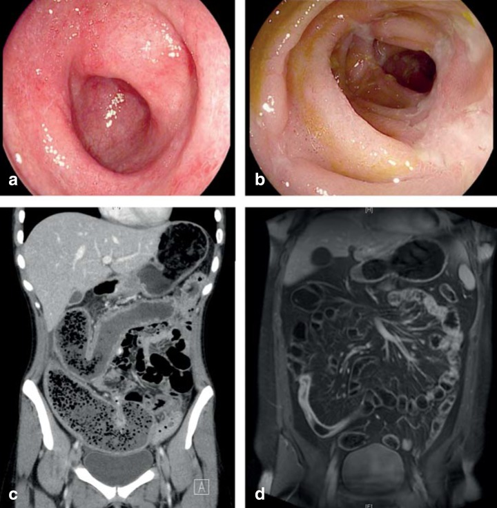
Figure 2.
Typical endoscopic and tomographic findings in chronic inflammatory bowel disease.
a) Mild ulcerative colitis: edema and erythema, with broken light reflexes from the granulated colonic mucosa.
b) Terminal ileitis in Crohn’s disease: partly aphthous (8 o’clock), partly confluent ulcerations (2 and 4 o’clock) of the small bowel mucosa.
c) Contrast-enhanced computerized tomography (CT): massively dilated cecum with coprostasis and thickening of the wall of the ascending and transverse colon with loss of haustration in an acute, severe relapse of longstanding ulcerative colitis (kindly provided by PD Dr. C. Schraml, Department of Diagnostic and Interventional Radiology [Director, Prof. Dr. K. Nikolaou], Universitätsklinikum Tübingen).
d) Contrast-enhanced magnetic resonance enteroclysis: Crohn’s disease, mixed inflammatory and fibrotic-stenotic changes in the terminal ileum (acknowledgement as above).

