Fig. 4.

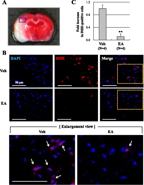
EA preconditioning reduced ROS production after focal cerebral ischemia. a The blue rectangle illustrates the imaging field. b Dihydroethidium (DHE, red, arrows) fluorescence images show the superoxide signal taken from the ischemic cortex 24 h after focal cerebral ischemia. The mice received EA (20 min) once a day for three days prior to the ischemic event. DAPI (blue), nucleus. Scale bar is 50 μm. c Quantification was done in three adjacent brain sections and three fields per predefined area of each brain section were counted for DHE staining in the ischemic cortex. Data are expressed as the means ± SEM (N = 4). ** P < 0.01 when compared with the vehicle group (Veh)
