
Fig. 2.
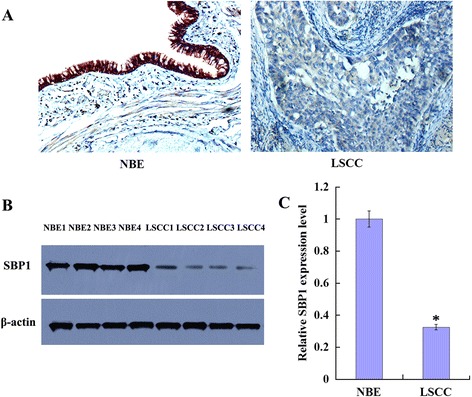
Expression of SBP1 in the human normal bronchial epithelium and lung squamous cell carcinoma tissues. a A representative result of immunohistochemistry shows expression of SBP1 is reduced in LSCC compared with the matched NBE. Original magnification, ×200. b A representative result of western blotting shows the expressions of SBP1 in the microdissected NBE and LSCC; c histogram shows the expression levels of SBP1 in NBE and LSCC tissues as determined by densitometric analysis. β-Actin is used as the internal loading control. Columns, mean from 16 cases of tissues; bars, SD (*P < 0.05 by one-way ANOVA)