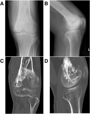
Fig. 1.

Plain X-ray findings and computed tomography findings. Plain X-ray (a, b) and computed tomography (c, d) show bone grafts, including a fibula graft performed at the time of initial surgery involving transplantation of the femoral metaphysis to the epiphyseal area. Neither bone translucency nor destruction is apparent on these images
