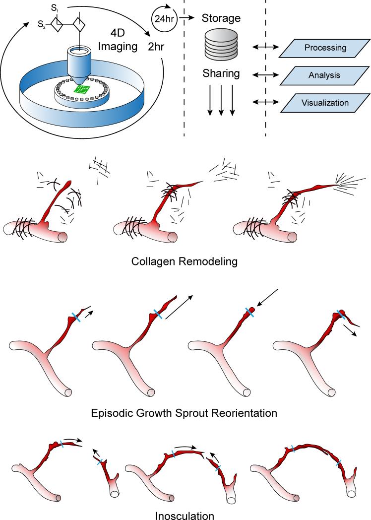
Figure 1.
Summary of the imaging and analysis protocol used in the study and the generalized findings related to neovessel activity. Top row: Schematic of the time-lapse acquisition involved an iterative process of twenty-five fields of view simultaneously recording SHG signals from collagen and fluorescence from EGFP in a mosaic fashion every 2 hours. Raw image data was transferred to an open-source storage server and processed off-line. Bottom Rows: Cartoons of the three main dynamic processes observed in the angiogenesis ex vivo system emphasizing the neovessel growth activities and collagen fibril restructuring associated with the growing neovessels.

