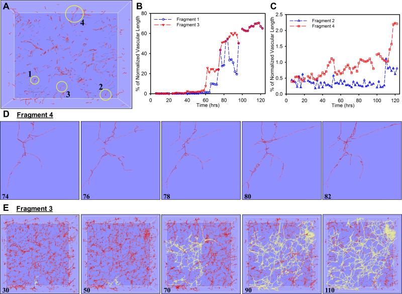
Figure 4.
Morphometric data for selected individual microvessel fragments as obtained from the skeletonized vascular networks. (A) - The percentage of total vascular length that was connected to an individual microvessel fragment was calculated for four random-selected initial fragments. (B-D) - vascular length percentage tended to oscillate over time as neovessels transitioned between periods of elongation and regression. Fragments 1 and 3 inosculated to the predominate vascular network forming within the construct which eventually accounted for roughly 70% of total vascular length (B, E), while Fragments 2 and 4 simply oscillated throughout time and did not experience any significant anastomosis events and remained unconnected to the majority of vascular perfusion within the construct (C).

