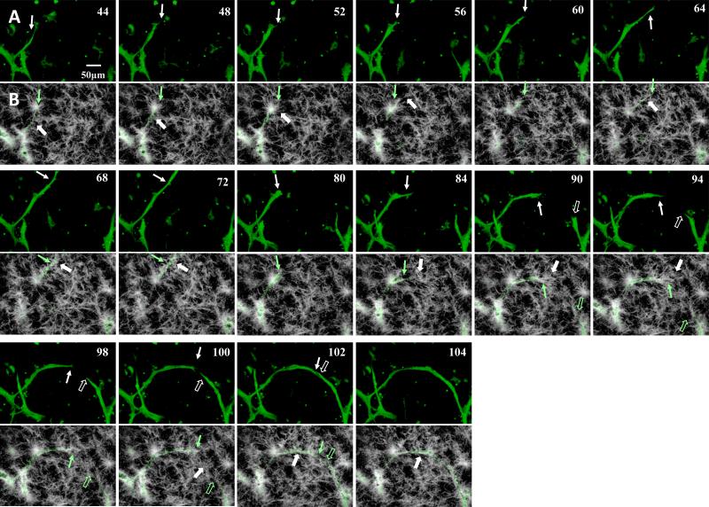
Figure 6.
A) Sequential (left-right, top-bottom) still-frames from time lapse movie of angiogenesis highlighting neovessel sprouting and growth activities via the EGFP channel for microvessel sprouts. Numbers in the top-right indicate acquisition time relative to the beginning of acquisition in hours. Notable features of the growing neovessel include sprout to body formation (44-52 hours), tip cell filopodia (60 and 64 hours), sprout advancement and retraction (72-80 hours), changes in growth direction (84 hours) and the directed growth towards and inosculation with adjacent neovessel sprouts (94-104 hours). While clearer in the suppl. movie 4, neovessel diameters fluctuated as the neovessel advanced, reflecting possible redistribution of cytoplasm. The solid and open arrows point to two different advancing neovessels. Representative time points were selected for this figure. B) - SHG channel of the time points illustrated in Fig. 6A. We only rendered frames over a depth of 10 microns at the sprout tip. The location of the sprout tip is illustrated with the green arrow. White arrows point to locations of collagen fibril recruitment, condensation or deposition. An aster-like fibril cluster served as stalk cell anchor (44-52). Fibrils were recruited behind the advancing sprout to surround it (68-72). Fibril reorientation occurred ahead of the advancing tip cell in frames 84 to 94. We observed unilateral condensation of fibrils at the stalk segment at the inside of the curved neovessel (102,104).

