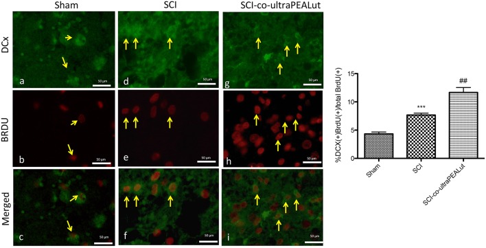
Figure 3.
Effect of co-ultraPEALut on co-localization of BrdU/DCX labeling after SCI. Results are shown for (A–C) sham-operated mice, (D–F) mice with SCI, and (G–I) mice with SCI treated with co-ultraPEALut. Spinal cord lesions sections, obtained from perilesioned area, were double-stained with antibodies against BrdU and DCX. The staining revealed that the increased proliferation of new neurons (DCX+ cells) started in mice subject to SCI (D–F, see yellow arrows that indicate the overlay) but treatment with co-ultraPEALut significantly increased the proliferartion after 72 h (G–I, see yellow arrows), see densitometric analysis. All images were digitalized at a resolution of 8 bits into an array of 2048 × 2048 pixels. (##p < 0.01 vs. SCI; ***p < 0.001 vs. sham).

