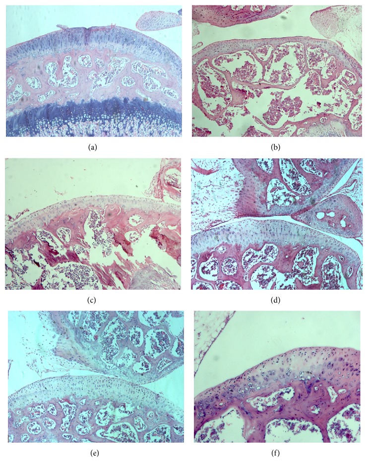
Figure 3.
HE staining of articular cartilage. Mature cartilage layer was recorded in normal mouse (a). Mature cartilage was thinned by needle (b). Injured cartilage was regenerated in negative control group (c, e) and treated group (d, f). However, the neocartilage in treated group was thicker than in negative control group.

