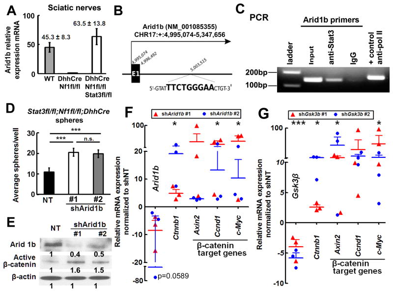
Figure 6. Stat3 transcriptionally represses Arid1b expression, activating β-catenin.
(A) qRT-PCR shows high Arid1b mRNA in Stat3fl/fl;Nf1fl/fl;DhhCre mouse sciatic nerve (white, n=6) versus WT (n=6) and Nf1fl/fl;DhhCre nerve (n=6). (B) Schematic, exon 1 mouse Arid1b gene. A putative Stat3 binding motif is between Exon 1 and Exon 2; the binding motif sequence is shown in bold. (C) Stat3 on the Arid1b promoter. PCR amplified a 139-bp Arid1b DNA fragment after ChIP with anti-Stat3. IgG was negative control. (D) Two Arid1b shRNAs increase numbers of Stat3fl/fl;Nf1fl/fl;DhhCre mouse neurofibroma spheres. (E) Western blot shows knockdown of Arid1b and increased β-catenin in Stat3fl/fl;Nf1fl/fl;DhhCre mouse neurofibroma spheres 4 days after shArid1b infection in two different shRNA clones. (F–G) β-catenin target gene expression increases after shArid1b (F) or shGsk3β (G) infection of Stat3fl/fl;Nf1fl/fl;DhhCre neurofibroma spheres. Mean ± SEM is shown for 3 independent experiments and 2 clones of shRNA in D, F and G. A representative experiment (of 3) is shown in C. Statistics: Ordinary one way ANOVA.

