Fig. 1.
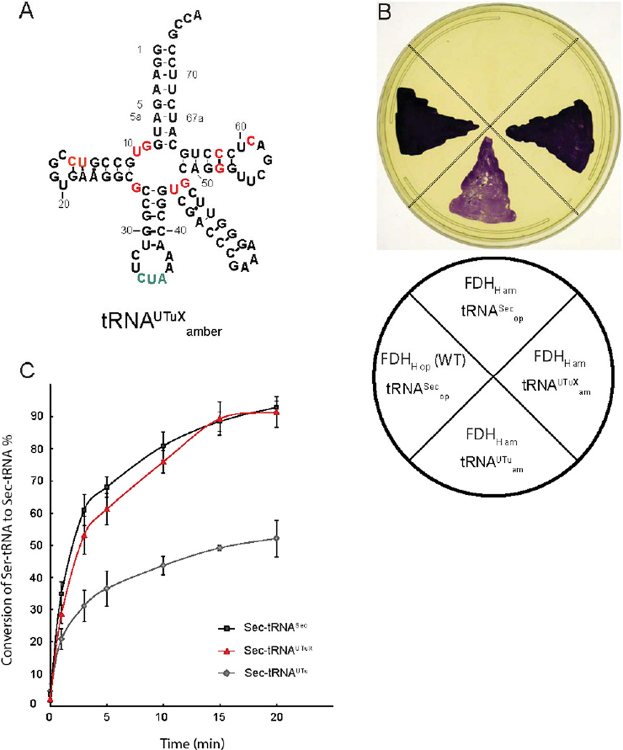
(A) Secondary structure of tRNAUTuX. Nucleotides that were changed from the original tRNAUTu are highlighted in red, the amber anticodon is depicted in green. (B) tRNAUTuX mediates functional Sec insertion in FDHH. An E. coli ΔselA ΔselB Δfdhf triple deletion strain was separately complemented with E. coli SelA, M. jannaschii PSTK alongside tRNASecop, E. coli SelB, and WT FDHHop; tRNAUTuam and FDHHam; tRNAUTuXam and FDHHam; and a negative control with tRNASecop, E. coli SelB, and FDHH140am. FDHH activity was assessed by appearance of the purple colored reduced BV. (C) In vitro conversion of Ser-tRNASec, Sert-RNAUTu, and Ser-tRNAUTuX by SelA. 5 µM SelD, Reactions were pre-incubated with 1 mM Na2SeO3 and 5 mM ATP at pH 7.2 under anaerobic conditions at 37°C for 30 min and then supplemented with 1 µM SelA and 10 µM of [α32-P] radiolabeled Ser-tRNA species for up to 20 min. Aliquots of 1.5 µL were taken at different time points, digested with nuclease P1, and spotted onto cellulose thin layer chromatography plates. After developing, plates were analyzed by autoradiography. While approximately 50% of Ser-tRNAUTu was converted, both tRNASec and tRNAUTuX support nearly full conversion to Sec-tRNA over a course of 20 min.

