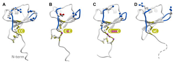
Figure 1.
Representative chemokines of CC (A), CXC(B), CX3C (C) and XC (D) families. The conserved cysteine pattern is shown in yellow sticks; residues separating the two N-terminal cysteines are shown as magenta balls (B, C). Loops believed to be involved in coordinating the N-terminus of the receptor (N-loop and 40s-loop) are colored blue and charged residues in these regions are shown as sticks.

