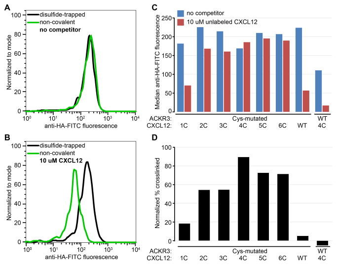
Figure 7.
Detection of crosslinked ACKR3:CXCL12 complexes by flow cytometry following incubation with an unlabeled competitor. (A) Due to high affinity and/or slow dissociation of the chemokine, anti-chemokine staining is indistinguishable between the non-covalent (green) and disulfide-trapped (black) complexes, (B) Prolonged incubation with excess unlabeled CXCL12 dissociates non-covalent (green) but not disulfide-trapped (black) complexes, effectively reducing surface staining. (C) Flow-cytometry screening of multiple disulfide-trapped candidate complexes side-by-side with controls (ACKR3(Cys):CXCL12 WT and ACKR3 WT:CXCL12(4C)). (D) Percent crosslinked for each complex was estimated by dividing median anti-HA-FITC fluorescence in the presence of competitor by that in the absence of competitor and subtracting non-specific signal from control samples.

