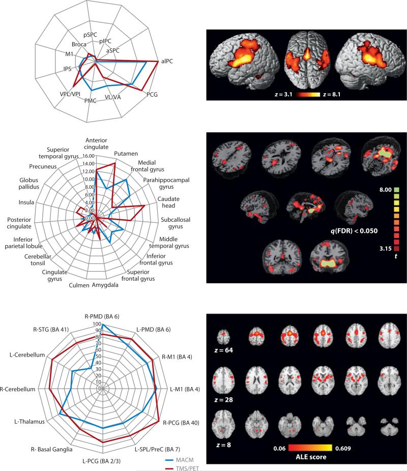
Figure 3.
Meta-analytic connectivity mapping (MACM) validations. The images illustrate three independent validations of MACM-derived connectivity patterns as forming one two-panel row. In each row, the left panel compares the strength of an MACM-derived projection with that of an alternative connectivity-mapping method. The right panels show the MACMs derived applying three different seed regions to the BrainMap database. The top row used DTI tractography as the validation methodology, seeding two regions within the parietal operculum (OP1, OP4; Eickhoff et al. 2010). The middle row used resting-state fMRI as the validation methodology, seeding nucleus accumbens (Cauda et al. 2011). The bottom row used concurrent transcranial magnetic stimulation (TMS)/PET as the validation methodology, seeding the supplementary motor area (SMA) (Narayana et al. 2012). Other abbreviations: aIPC, anterior inferior parietal cortex; aIPS, anterior intraparietal sulcus; aSPC, anterior superior parietal cortex; Broca, Broca's area; L-M1, left primary motor cortex; L-PCG/BA2/3, left postcentral gyrus, Brodmann areas 2 and 3; L-PMD, left dorsal premotor cortex; L-SPL/PreC, left superior parietal lobule/precuneus; M1, primary motor cortex; PCG, postcentral gyrus; pIPC, posterior inferior parietal cortex; PMC, premotor cortex; pSPC, posterior superior parietal cortex; R-M1, right primary motor cortex; R-PCG, right postcentral gyrus; R-PMD, right dorsal premotor cortex; R-STG (BA 4), right superior temporal gyrus, Brodmann area 41; VL/VA, ventrolateral nuclei/ventrolateral anterior nuclei; VPL/VPI, ventroposterior lateral and inferior nuclei. Images are reproduced with permission from each manuscript.

