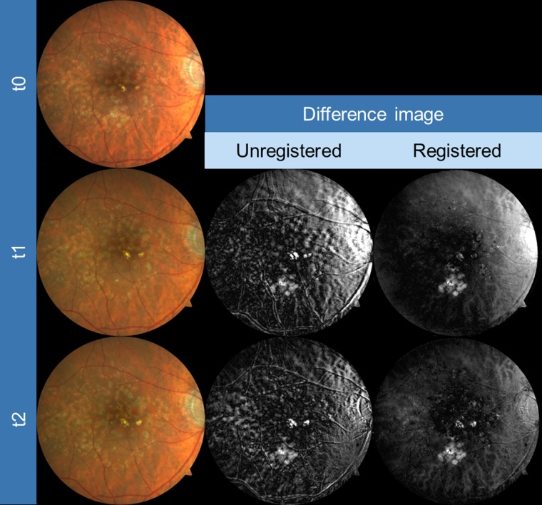
Figure 1.
Exemplary images of an eye for CFP registration as obtained from three different time points (baseline [t0], year 1 [t1], year 2 [t2], first column). Slight but obvious misalignment in the unregistered images is visible, particularly when focusing on retinal blood vessels (gray-scale difference of baseline and both t1 and t2 images, second column). Automated registration clearly improved the alignment (third column).

