FIGURE 3.

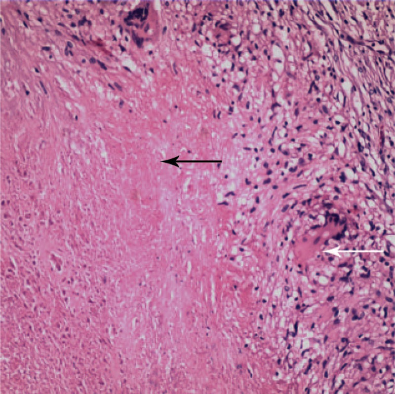
Histopathological examination of the left supraclavicular lymph node showed caseous necrosis (black arrow) and multinucleated giant cells (white arrow). The final diagnosis was lymph node tuberculosis.

Histopathological examination of the left supraclavicular lymph node showed caseous necrosis (black arrow) and multinucleated giant cells (white arrow). The final diagnosis was lymph node tuberculosis.