
Figure 4.
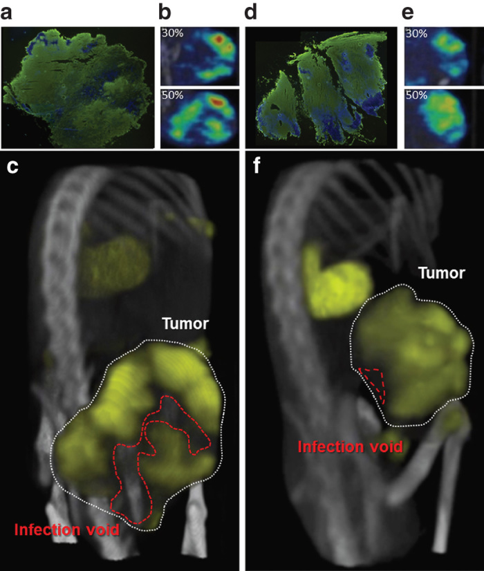
Space filling models show infection voids. The need for three-dimensional (3D) imaging is made clear when comparing tumors from multiple animals. (a/d) Immunohistochemical staining of tumors shows extensive vesicular stomatitis virus (VSV) infection (anti-VSV, green) throughout the tumor (Hoechst, blue). (b/e) Singular microSPECT/computed tomography (CT) planes taken at the same relative position within the same tumors as those stained in a/d show similar distribution relative to each other (top) but different planes through the same tumors at another location show different distributions. (c/f) 3D space filling allows differences in distribution to be clearly appreciated where tumor on top has decreased voids compared to tumor on bottom.