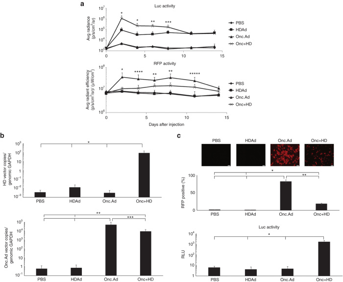
Figure 4.
Coinfection of Onc.Ad amplified HDAd vector DNA and transgene product following intratumoral injection. (a) A549 cells were transplanted subcutaneously into the right flanks of nude mice, and a total of 1 × 10⁸ Vp of Onc.Ad alone, HDAd alone or an Onc.Ad/HDAd mix (Onc.Ad to HDAd; 1:20) was injected intratumorally. Luciferase (HDAd) and RFP (Onc Ad) activity in tumors were measured at 0, 2, 4, 6, 8, 11, and 14 days after injection. Data are presented as means ± SD (n = 6). The experiment was repeated twice with similar results. *P < 0.001, **P < 0.004, ***P < 0.01, ****P < 0.002, *****P < 0.03. (b) Tumors were collected and harvested at 15 days after injection. DNA was extracted from each tumor sample, and the copy number of each vector determined by quantitative PCR using primer sets for each vector backbone. Data was normalized with human genomic GAPDH. Data are presented as means ± SD (n = 6). *P < 0.001, **P < 0.03, ***P < 0.01. (c) Homogenized tumor samples were lysed via 3 freeze-thaw cycles, and a portion of the tumor lysate was added to untreated A549 cells in vitro. RFP fluorescence (Onc Ad) was captured at 24 hours after treatment, and the population of RFP positive cells were analyzed by Flow cytometry. The luciferase activity (HDAd) of cells treated with tumor lysates was also measured at 24 hours after treatment. Data are presented as means ± SD (n = 6). *P < 0.003, **P < 0.001.

