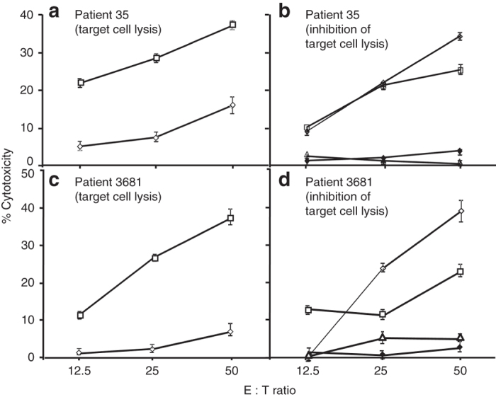
Figure 6.

Cytotoxic activity of HLA-A2 peptide-stimulated PBMC of HLA-A2+ melanoma patients against target cells (a–d). Short-term T-cell lines derived from PBMC of HLA-A2+ melanoma patients after peptide-pulsed monocytes and IL-2 stimulation in vitro as in Figure 5 were tested for cytolytic activity against HLA-A2+ target (WM35) cells in a 51Cr release assay. PBMC from patient 35 (a) and patient 3681 (b) were stimulated with either peptide A2_1 (◊) or A2_2 (□) and were tested for cytotoxic activity against 51Cr-labeled WM35 tumor cells as targets. To determine MHC restriction of peptide A2_2 stimulated PBMC (35 PBMC (b); 3681 PBMC (d)), 51Cr release assay was performed in the presence of medium (◊), normal mouse IgG (□), anti-HLA class I antibody (∆) or anti-HLA-A2 antibody (♦). Results are expressed as mean % cytoxicity ± SE (a–d) and cytotoxic T lymphocyte lysis of WM35 (HLA-A2+) target cells was significantly inhibited (P < 0.001) in presence of anti-HLA class I or anti-HLA-A2 antibodies. All experiments were repeated at least twice for confirmation.
