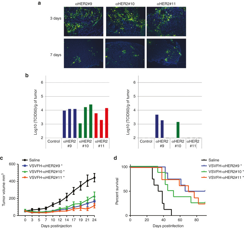
Figure 5.
Antitumor efficacy of VSV-FH and HER2-targeted viruses. Subcutaneous SKOV3ip.1 tumors were implanted in the right flank of nude NCr mice. Once tumors reached a volume of 50 mm3, 1 × 10⁶ TCID50 units of VSVFH-αHER2 (αHER2) #9-11 were intratumorally administrated in 50 µl of OptiMEM. (a) VSV-positive cells at 3 or 7 days post-treatment were detected by immunofluorescence using an anti-VSV polyclonal antibody. (b) Number of infectious particles present in the tumors, at the same days post-treatment, were determined in the tumors of three mice treated with the HER2-targeted viruses. (c) Tumor growth was monitored three times per week. Average of the tumor size of the mice per group (n = 8) was plotted plus standard error of the mean. (d) Survival of mice treated with the indicated viruses or saline (control). Asterisk (*) indicates the groups that are statistically different from saline control group.

