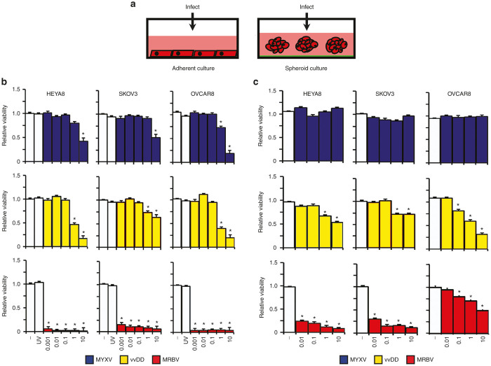
Figure 2.
Analysis of MYXV, vvDD, and MRBV oncolytic-mediated killing of EOC cell lines in adherent and spheroid culture. (a) Schematic representation of viral infection of ovarian cancer cells in adherent and spheroid culture. (b) HEYA8, SKOV3, and OVCAR8 cells were infected at increasing concentrations to a maximum of multiplicity of infection (MOI) 10, as indicated; UV-inactivated virus was used at a MOI of 10. Cell viability was measured after 72 hours using CellTiter-Glo. (c) HEYA8, SKOV3, and OVCAR8 cells were seeded to Ultra-Low Attachment dishes to form spheroids over 3 days, then infected at the indicated doses; spheroid cell viability was assayed as in panel b (*P < 0.05). EOC, epithelial ovarian cancer; MYXV, Myxoma virus.

