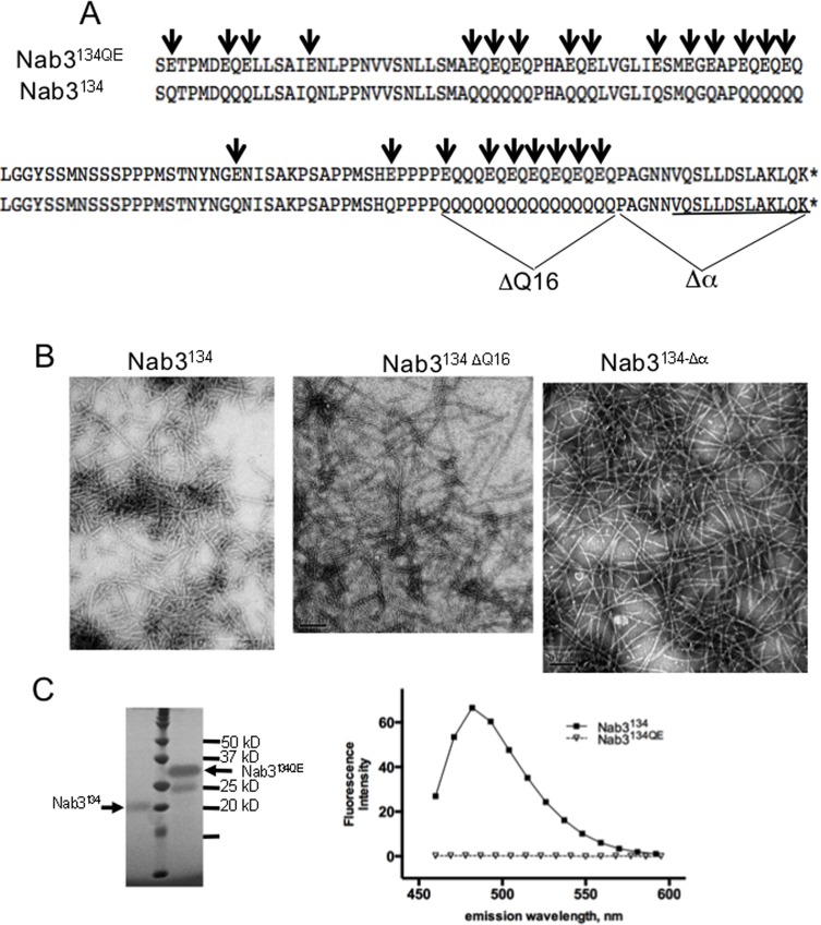
Fig 1. Filament formation by mutants of the LCD of Nab3.
(A) A portion of the wildtype (Nab3134) and glutamine to glutamate substituted (Nab3134QE) LCD of S. cerevisiae Nab3 is shown. The extent of the deletion of Q16 run and the terminal hnRNP-C-like domain (Δα) are also shown. The region of Nab3 with structural homology to human hnRNP-C is underlined. (B) Transmission electron microscopy images of filaments from the various purified versions of Nab3134 are shown. (C) Purified Nab3134 and Nab3134QE were run on a 12% SDS-PAGE gel (left panel) next to molecular weight markers and stained with ImperialTM stain (note this polypeptide binds Coomassie blue-based dyes poorly). Thioflavin T-binding and fluorescence (right panel) was measured for Nab3134 and Nab3134QE as described in Materials and Methods.

