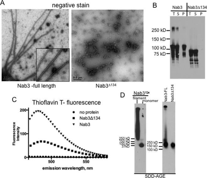
Fig 2. Amyloid formation by full-length Nab3 protein.
Purified Nab3 and Nab3Δ134 were examined by transmission electron microscopy (A), or pelleted and subjected to Western blotting (B). Total (T), supernatant (S), and pellet (P) fractions were analyzed in (B). Nab3 and Nab3Δ134 were incubated with thioflavin T and examined by spectrofluorimetry at the indicated wavelengths (C) or subjected to SDD-AGE using anti-Nab3 antibody (D; right panel). As a control, Nab3134 filaments and monomers were analyzed on a separate gel with antibody against S-tag (left panel) by SDD-AGE.

