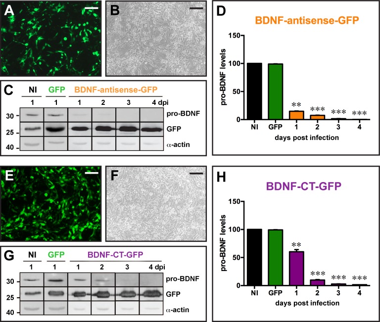
Fig 2. In vitro validation of the amplicon vectors.
(A to D) Infection of mesoangioblast cells (MABs) constitutively expressing BDNF with the BDNF-antisense-GFP amplicon vector at MOI 5. Infection of the cells with amplicon vectors was confirmed by GFP fluorescence (A) and GFP detection on western blot (C). Pro-BDNF expression was analyzed by western blot in the 4 days following infection and pro-BDNF signal was normalized to α-actin for quantification (D). (E to H) Infection of MABs with the BDNF-CT-GFP amplicon vector at MOI 5. Infection of the cells was confirmed by GFP fluorescence (E) and GFP detection on western blot (G). Pro-BDNF expression was analyzed by western blot in the 4 days following infection and pro-BDNF signal was normalized to α-actin for quantification (H). Data in D and H are the mean±SEM of 6 experiments. * p<0.05, **p<0.01, ***p<0.001: ANOVA and post-hoc Dunnett test. Horizontal bars in panels A, B, E and F = 25 μm.

