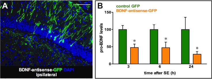
Fig 4. Transgene expression following injection of amplicon vectors in the right and left hippocampus at different time points after pilocarpine-induced status epilepticus.
(A) Representative GFP immunofluorescence in the dorsal hippocampus of a rat at 5 days post injection with the BDNF-antisense-GFP amplicon vector. (B) Quantification of the pro-BDNF signal, normalized to α-actin, 3, 6 and 24 h after pilocarpine status epilepticus induced 5 days after injection of the amplicon vectors in the right dorsal hippocampus. Data in B are the mean±SEM of 4–5 rats per group. * p<0.05, ANOVA and post-hoc Dunnett test. Horizontal bar in A = 250 μm.

