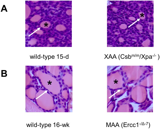
Fig 3. Histological examination of haematoxylin/eosin-stained thyroid glands of 15-day-old WT and XAA (Csbm/m/Xpa-/-) (A) and 16-week-old WT and MAA (Ercc1-/Δ-7) (B) mice (all magnifications 10x).
The thyroid follicles (denoted by asterisk) surrounded by thyrocytes (denoted by arrow) are similar between WT and progeria models.

