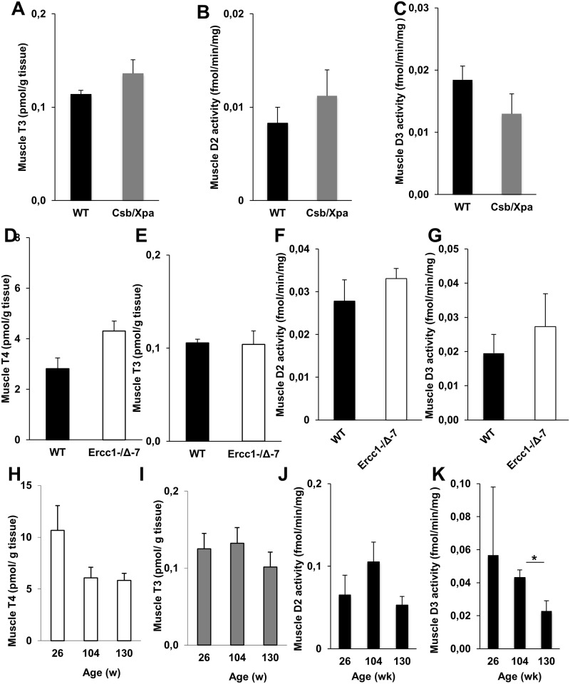
Fig 6. Thyroid state in skeletal muscle of progeroid and naturally aged mice.
T3 concentrations (A) and activities of D2 (B) and D3 (C) in muscle of 15-day-old WT and XAA (Csbm/m/Xpa-/-) mice (n = 3/group). T4 (D) and T3 (E) concentrations and activities of D2 (F) and D3 (G) in muscle of 18-week-old WT and MAA (Ercc1-/Δ-7) mice (n = 3/group). T4 (H) and T3 (I) concentrations and activities of D2 (J) and D3 (K) in muscle of 26-, 104-, and 130-week-old WT mice (n = 3-5/group). Values represent mean ± SE per group. * P < 0.05

