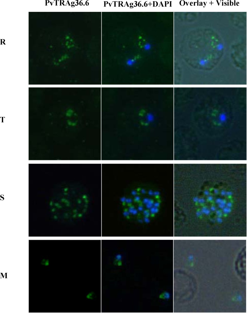
Fig 2. Sub cellular localization of PvTRAg36.6 in P.vivax natural infections.
Immunofluorescence images of P.vivax infected red cells. Parasites were labeled with anti- PvTRAg36.6 (green) antibody and DAPI for nuclear staining (blue). Fluorescence pattern observed in ring (R, double infection), trophozoite (T), Schizont (S) stages and in free merozoites (M) are shown. Overlay shows the images merged with bright field.

