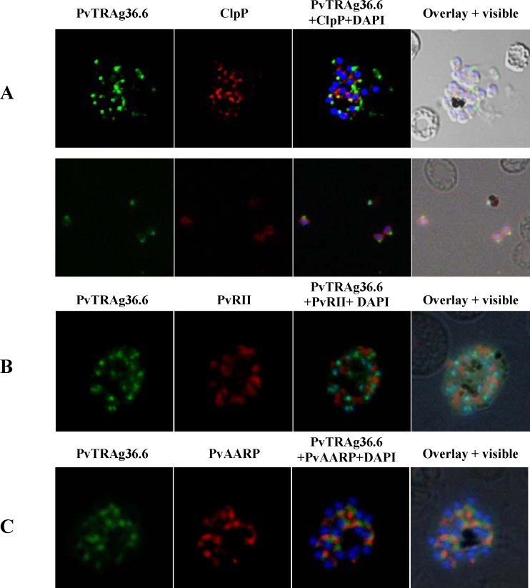
Fig 3. Co-localization studies of PvTRAg36.6 in P.vivax.
Co-localization images of PvTRAg36.6 with apicoplast, rhoptry and micronemal markers in P.vivax natural infections. (A) Fluorescence pattern observed after co-immuno staining of P.vivax parasite with anti-PvTRAg36.6 (green) and anti-PfClpP (red) recognizing apicoplast in schizont (A, upper panel) as well as in free merozites (A, lower panel). (B) Co-immunostaining of anti-PvTRAg36.6 (green) with anti-PvRII (red) recognizing microneme in a schizont, and (C) Co-immunostaining of anti-PvTRAg36.6 (green) with anti-PvAARP (red) recognizing rhoptry neck in a schizont. The parasite nuclei were stained with DAPI (blue). Overlay shows the images merged with bright field.

